


Veterinarians often need to see inside a sick or injured dog to figure out what’s wrong. But what’s the best imaging technique to use? It depends on what the veterinarian suspects might be the problem. What follows is a guide to the various ways doctors view what’s happening under the coat. As you’ll see, the cost of imaging is a good reason to have pet health insurance.
X-ray
Detects: Major abnormalities and dramatic changes in the size, shape, or content of organs.
How it works: Differentiates between bone, gas, soft tissue, fluid, and fat because various internal organs and bone absorb electromagnetic radiation (x-rays) in differing amounts. Bone looks whiter than the rest of the image because it absorbs more x-rays. Gas looks black because it absorbs none.
Sedation necessary? Not usually (which helps keep down cost).
Cost: (roughly) $50 – $100 per image.
X-rays are used most often in dogs for images of the chest or abdominal cavity. They help diagnose lung and heart disease and can also help determine the reason for unexplained vomiting or diarrhea. They’re good, too, for diagnosing bone fractures or lameness caused by a joint problem.
Ultrasound
Detects: Most body tissues and their architecture, including the heart, abdominal organs like the stomach and liver, and muscles and tendons.
How it works: Sound waves transmitted into the patient are reflected back from tissue based on its acoustic properties — its ability to transmit sound. (Bone and gas do not allow the transmission of sound waves, so ultrasound is not good for looking at them.)
Sedation necessary? Not usually.
Cost: (roughly) $300 – $400.
Ultrasound allows the veterinarian to see detail inside an organ that cannot be seen on an x-ray. For example, it can allow the blood supply to the organ to be assessed, or it can allow a small tumor within an organ to be visualized. In addition, ultrasound allows organs to be observed over a several-minute period: ultrasound is a “movie,” while an x-ray is a “snapshot.”
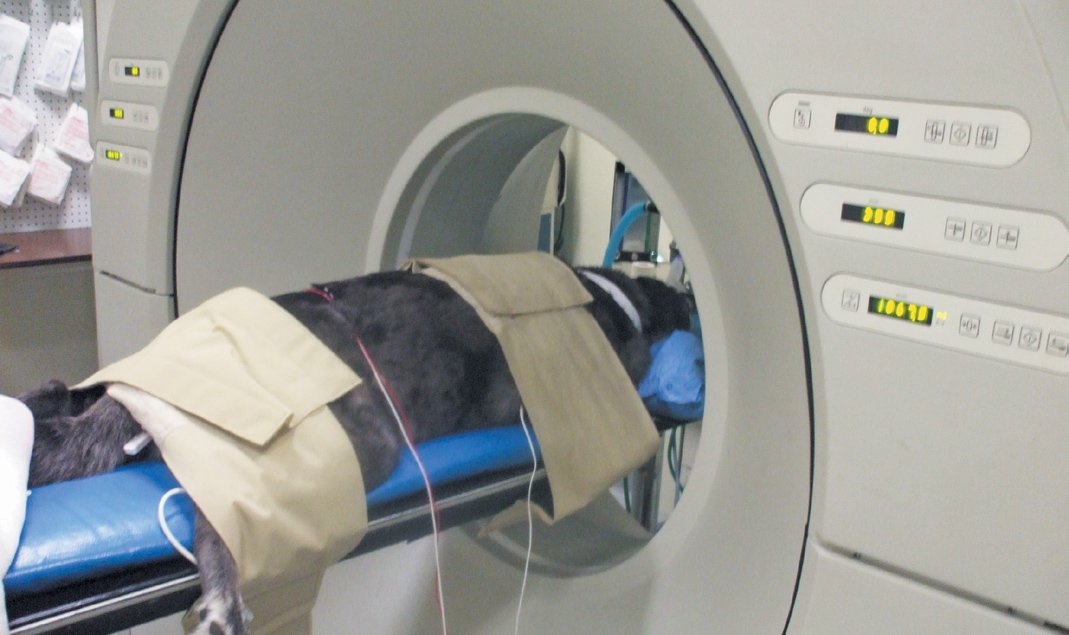
CT Scan
Detects: CT scans can be used to diagnose diseases almost anywhere in the body, but they are used most frequently for the head, neck, chest, abdomen, and spine.
How it works: A CT scanner takes large numbers of individual x-rays, then uses a computer to reconstruct these images into a three-dimensional view deep inside the body rather than just a flat, two-dimensional view. The technology allows the interior of the body to be viewed from multiple perspectives.
Sedation necessary? Yes, most often in the form of general anesthesia.
Cost: (roughly) $500 – $1,500 depending on the complexity of the study (plus a few hundreds dollars for the anesthesia).
MRI
Detects: Any type of tissue disease or injury, although most commonly applied to the brain and spinal cord.
How it works: MRI (magnetic resonance imaging) uses hydrogen atoms to obtain image contrast. A strong magnetic field temporarily aligns the body’s hydrogen atoms. A radio frequency current then manipulates the hydrogen atoms to produce signals that create a cross-sectional picture.
Sedation necessary? Yes. It often takes more than an hour to complete the imaging — far longer than any dog would be able to lie perfectly still.
Cost: (roughly) $1,100 to $1,500 (plus a few hundred dollars for the anesthesia).
MRI can identify very subtle problems that wouldn’t necessarily show up on a CT scan. And it picks up brain pathology very well because most diseases, such as tumors and inflammatory diseases, have increased water content and thus a high concentration of hydrogen atoms.
Nuclear Medicine
Detects: Alterations in how an organ or body system is functioning rather than simply what it looks like.
How it works: Cells or drugs “tagged” with radioactivity are administered to the patient, allowing the radiologist to see how these substances move through or interact with organs.
Sedation necessary? Yes. Short-acting sedatives can be used instead of full-on anesthesia.
Cost: (roughly) $300 – $400 (plus $100 to $200 for the sedation, less than full anesthesia).
Veterinary radiologists use nuclear medicine to trace where blood is leaking during instances of internal hemorrhaging. This technique can also assess the kidneys’ filtering ability, and can be used to identify metastases of tumors to bones. But it is probably most often used to diagnose congenital blood vessel abnormalities in the abdomen.


