


Rocky was rushed to Tufts’s small animal hospital in the middle of the night. The 2-year-old Brittany spaniel was coping with extreme discomfort and dehydration and had now started vomiting. To find out quickly what was wrong — maybe Rocky had an intestinal blockage that needed to be repaired immediately so he wouldn’t die — the emergency veterinarian had to decide what kind of picture to get of his gastrointestinal tract, where the problem was suspected. Would the young dog be best served by a set of x-rays, which would cost in the neighborhood of $100 to $200, or would he need, say, an ultrasound, which would perhaps triple or quadruple the cost?
Everybody has heard of x-rays, ultrasounds, CT scans, and other types of medical imaging — most of us have had at least one of them ourselves at one point or another. But few know exactly what each type of image captures, and when each is called for. Here’s a run-down.
X-Rays for Dogs
Best for seeing: major abnormalities and dramatic changes in the size, shape, or content of organs.
Picks up: bone; gas; soft tissue and fluid; fat; metal.
How it works: Different tissues absorb x-rays (electromagnetic radiation) in differing amounts. Bone will look whiter than the rest of the image because it absorbs more x-rays. Conversely, gas will look black, as it absorbs no x-rays. An organ such as the spleen will come across gray.
Sedation necessary? Not usually (which helps keep down cost). Exceptions include anxious or boisterous dogs or those in pain.
Cost: (roughly) $50 to 100 per image. X-rays are used in dogs most often for images of the thoracic (chest) cavity and the abdominal cavity. “In the thorax we’re looking for lung and heart disease, predominantly,” says Tufts board certified veterinary radiologist James Sutherland-Smith, BVSc, DACVR. “Coughing, struggling to breathe, or breathing too fast might be what leads to a thoracic x-ray. An abdominal x-ray might be triggered by unexplained vomiting or diarrhea. Occasionally something like struggling to urinate may be a reason to x-ray inside the abdominal cavity, too.” Most of the gastrointestinal organs are housed in the abdomen.
X-rays are also good for helping to diagnose bone disease such as a fracture or joint problem that might be causing lameness. And they can be very useful in picking up internal damage after a trauma such as a dog’s getting hit by a car. Dr. Sutherland-Smith says, “you can see the bruising of a lung or a torn diaphragm” — a sheet of muscle that is supposed to keep the chest and the abdomen separate.
One advantage of an x-ray is that the dog can typically be fully awake for the procedure. “We use positioning devices like sandbags that we wrap around a dog’s legs,” Dr. Sutherland explains. “And we have ‘bookends’ — padded heavy weighted metal devices that we can use to hold a dog in position. Small dogs — we’ll have someone actually hold them. The person is gowned up with a lead apron.”
X-rays’ major limitation is their inability to distinguish more than four main densities in the normal body. For example, an x-ray can tell bone from soft tissue and fluid, but it can’t tell what soft tissue or fluid you’re looking at. It will all come out the same shade of gray. “The liver and spleen look identical” on an x-ray, Dr. Sutherland-Smith says. “The fact that we determine it’s the liver is based on location. There’s some guesswork, or interpretation, involved.” Also, while an x-ray can show that the liver is, say, twice its normal size, “it’s poorly sensitive to more subtle things,” Dr. Sutherland-Smith points out. “I can’t see if it has a change within the tissue such as a small nodule” that might be a tumor.
Ultrasounds for Dogs
Best for seeing: most body tissues and their architecture, including the heart, abdominal organs, and musculoskeletal system (muscles and tendons).
How it works: Sound waves are transmitted into the patient and reflected back from tissue based on the architecture and acoustic properties of that specific tissue. (Ultrasound is not good for looking at bone or gas-filled lungs, both of which do not allow the transmission of sound waves.)
Sedation necessary? Not usually (which helps keep down cost). Exceptions include anxious or boisterous dogs or those in pain.
Cost: (roughly) $300-$400.
With an ultrasound, unlike an x-ray, you can “discriminate between different things going on in one organ,” Dr. Sutherland-Smith says. “It won’t be just all one shade of gray.” For instance, if you’re looking at the liver and there’s a tumor nodule, the sound waves will reflect that differently, and it will come across as a different color and therefore get picked up.
An ultrasound also looks at the workings of tissue in real time. It’s not just a snapshot of a single moment. For instance, you can see blood flowing through the chambers of the heart. You can see if any of the valves don’t close properly when blood goes through them and thus cause some blood to flow backward instead of forward, like it should.
You can even see differences in fluid types you’re zeroing in on, Dr. Sutherland-Smith says. For instance, if you’re looking at the bladder, abnormal urine (perhaps containing blood or crystals) will be more reflective than normal, and it will come across as a white or gray dot in a sea of black. You could also see “cellular debris if there were an infection in the bladder, like cystitis,” Dr. Sutherland-Smith explains.
Ninety percent of ultrasounds performed on dogs is for imaging of structures in the abdominal cavity or for looking at the heart, Dr. Sutherland-Smith points out. Occasionally, he says, ultrasounds will also be used to look at musculoskeletal issues — tendons and ligaments around the joints — or abnormal structures within the thoracic cavity.
The dog does not usually have to be sedated unless she is particularly excitable or nervous. On the day we visited the ultrasound unit on the radiology floor of Tufts’s Foster Hospital for Small Animals, a dog was lying on her back, gel smeared across her belly, for a look with an ultrasound probe to see if her mast cell cancer had metastasized to any body organs. The dog was as comfortable as could be, with several technicians at her side to help her feel secure.
One difference between abdominal ultrasounds for people and those for dogs is that with dogs, the medical team will generally look at all of the organ systems at one time rather than just in one location. “If the owner is spending a lot of money, we want to make sure he or she is not missing something else that will change the treatment plan,” Dr. Sutherland-Smith says. “By the time we perform an abdominal ultrasound, an older pet can often have more than one disease, and some of the illnesses may not have any outward signs for the owner or veterinarian to notice.”
Note: Sometimes, whether to order an abdominal ultrasound or an x-ray is a judgment call. “If it’s not urgent,” Dr. Sutherland-Smith says, meaning the dog is stable and not in acute pain, “most people will wait to do an ultrasound” as the procedure cannot generally be performed on the spur of the moment. But if the dog is in bad shape, an x-ray might be ordered to see if something has ruptured, torn, or broken, or if there’s a life-threatening blockage in the G.I. tract that needs immediate surgery. (At Tufts and some other state-of-the-art veterinary hospitals, both x-rays and an ultrasound can be had on an emergency basis.)

CT Scans on Dogs
Best for seeing: diseases of the head, including trauma to the head and nasal diseases, in addition to lung diseases (although CT scans can be used to create images of any part of the body).
How it works: CT scans use the same basic principle as x-rays. They are x-rays, in fact. But the difference is that they use more advanced technology to be able to get a three-dimensional view rather than just a flat, two-dimensional view. Think of a loaf of bread, Dr. Sutherland-Smith says. An x-ray can show you the loaf from the top, sides, or bottom. But a CT scan gives you cross-sectional imaging so that you can look at each slice of the loaf — even “pick it up and then put it back,” he says. It’s computed tomography (CT) that allows for the 3-D view. A body organ or tissue is scanned with x-rays, and a computer is used to construct a series of cross-sectional scans that “build” a full image. (Doctors who have separated conjoined human twins have used CT scans to build stand-alone models of how the two babies are connected so they can figure out how to release them from each other without harming critical blood vessels and various tissues. And our own veterinary orthopedic surgeons have used CT data to get models of dog bones that either are malformed or have had fractures that healed with excessive angulation, allowing them to practice complicated reconstructive surgeries.)
Sedation necessary? Yes. In fact, with rare exceptions, CT scanning requires general anesthesia. The dog must be kept still for several minutes while inside the scanner, which looks like a donut. It’s the spinning of the x-ray machine inside the “donut” that allows enough pictures of the tissue or organ to be taken in order to build a 3-D image.
Cost: (roughly) $500-1200 depending on the complexity of the study (plus a few hundred dollars for the anesthesia).
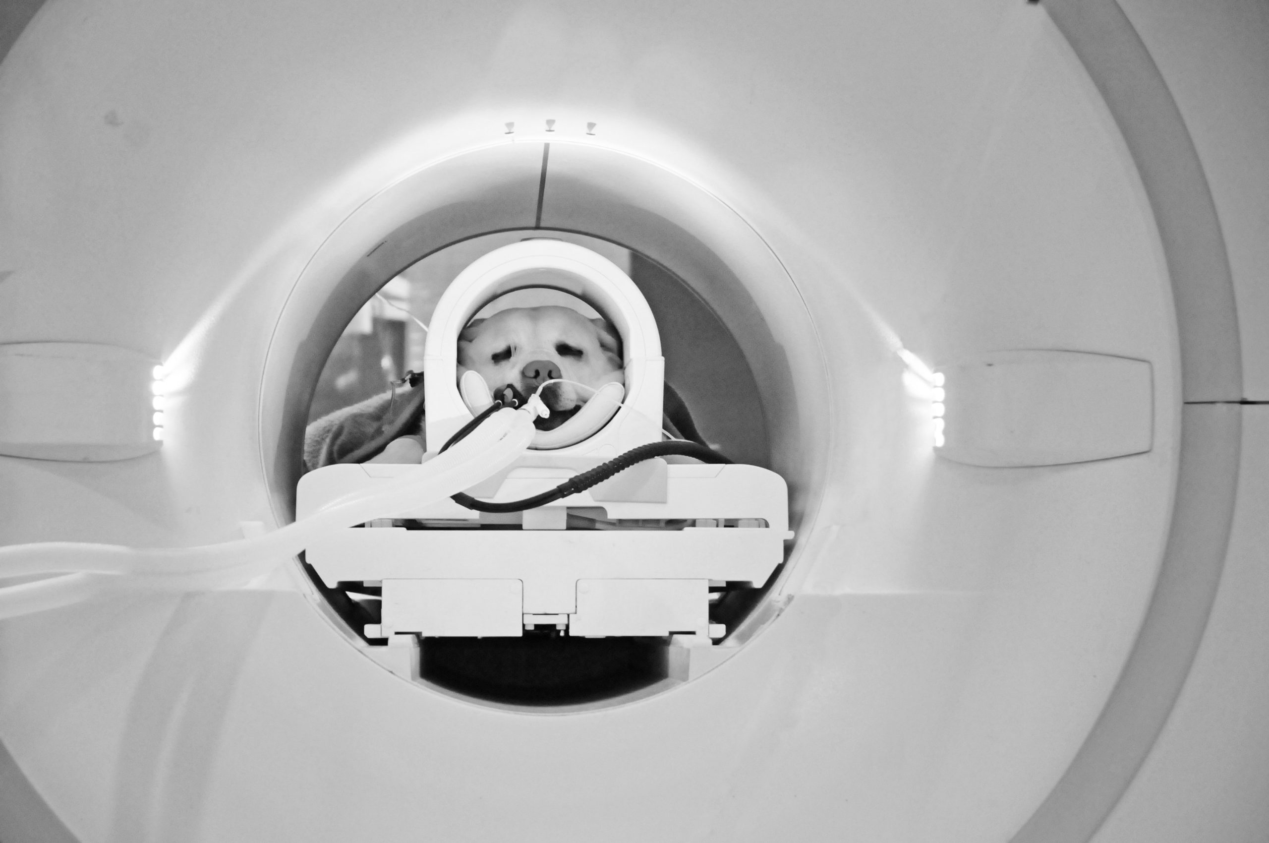
When Dogs Need MRI
Best for seeing: any type of tissue disease or injury, although most commonly applied to the brain and spinal cord.How it works: MRI (magnetic resonance imaging) uses the stimulation of hydrogen atoms to get contrast in its images. A very strong magnetic field temporarily aligns the mobile hydrogen atoms in the body. Radio waves then manipulate these atoms to produce signals that are used to create cross-sectional images — as with CT scans, like slices in a loaf of bread.
Anesthesia necessary? Yes. A dog must remain very still in the donut-shaped MRI machine, just like in the CT scan apparatus. Unlike CT, it often takes more than an hour to complete the imaging — far longer than any dog would be able to lie perfectly still.
Cost: (roughly) $1,100 to $1,500 (plus a few hundred dollars for the anesthesia).
“Probably 90 percent of what we do with MRI is neurologic imaging,” says Dr. Sutherland-Smith, that is, “imaging of the brain and spine. MRI can identify quite subtle pathology that wouldn’t necessarily show up on a CT scan.”
Another way of putting it: MRI is very good at differentiating mild differences in tissue; “it picks up disease very well,” Dr. Sutherland-Smith says, because most diseases and tumors and inflammation in the brain have increased water content and therefore a high concentration of mobile hydrogen atoms.
To take a recent case, an 11-year-old Lab mix came to Tufts falling over and turning his head to the left. Clearly, it looked like a neurologic problem originating in the brain. An MRI showed that he had a very large tumor of his skull between his ears — some 3 inches by 2 inches, which is quite large for a dog. (Amazingly, for a tumor of this size there was scant evidence of a bump on the dog’s head.) The tumor was going to be biopsied to see if it was cancerous, but whether benign or malignant, a tumor growing next to the brain is not okay. It can push in on adjacent areas of the brain and affect function, which is exactly what was happening. The tumor was pressing on a part of the dog’s brain that controlled balance, and needed to be treated.
Nuclear Medicine for Dogs
Best for seeing: information on how an organ or a system is functioning rather than what it looks like per se.
How it works: Cells or specific drugs are “tagged” with radioactivity so it can be seen how they move through or are processed by organs. The veterinarian can use specific molecules for tagging, depending on whether he wants to look at, say, kidney functioning or the path of red blood cells if internal bleeding is suspected.
Sedation necessary? Yes — short-acting sedatives are used.
Cost: (roughly) $300 to $400 (plus $100 to $200 for the sedation — less expensive than full anesthesia).
One thing nuclear medicine allows veterinarians to do is “follow” internal hemorrhaging. “We can label red blood cells with a very small dose of radioactivity and then image them — see how they get distributed through the dog and, hopefully, find an area where they’re leaking into the intestinal tract, kidney, or lower urinary tract,” Dr. Sutherland-Smith says. “Of course, this would be used only for a more challenging case of internal bleeding.”
Nuclear medicine can also assess the filtering abilities of the kidneys by attaching radioactivity to a drug that “gets cleared through the kidney,” the doctor comments. “We might use it if there’s a tumor in a kidney that requires removal of that kidney. We want to make sure the other one is functioning well enough to compensate for the one that’s going to be gone.”
But “probably the most common reason we do nuclear medicine in dogs,” Dr. Sutherland-Smith says, “is to look at congenital blood vessel abnormalities in the abdomen.” The most common of these is called a portosystemic shunt, which tends to occur in small dogs and toy breeds. Blood is supposed to go through the liver so that organ can filter toxins, but instead it goes straight to the heart — and from there to the brain, where it causes neurologic problems. This type of shunt can be fixed but can be hard to find. Nuclear medicine helps by proving that a shunt is present, and it will usually show an approximate path of the abnormal blood flow. “If we know that,” Dr. Sutherland-Smith says, “we can correct the problem surgically.”
Rocky, the dog mentioned at the beginning of this article, didn’t have any blood vessel abnormalities. In fact, as his x-ray showed, he didn’t even have excess gas — his intestine wasn’t bulging in any spot — and certainly no blockage or other life-threatening problem. He was simply given some fluid under his skin for his mild dehydration, along with an antacid, and was sent on his way. Sometimes imaging turns out not to show much of a problem at all.


Thanks for all the information. Good to know in advance of a real need.
Where can I find an affordable MRI for a dog?
My dog needs an MRI, however it is extremely expensive. Where do they offer low cost MRI’S?
I keep listening to the news lecture about getting free online grant applications so I have been looking around for the top site to get one. Could you advise me please, where could i acquire some?